Advanced Imaging: Crucial for Pet Health
Caring Hands Animal Hospital utilizes both radiology and ultrasound as essential diagnostic tools to support your pet's care. Ultrasound, or sonography, employs sound waves emitted from a probe, which are then transformed into visual images. These images are displayed on a screen, providing the veterinarian with a two-dimensional view of your pet's internal organs.
Radiology, or X-ray imaging, is a valuable diagnostic method for visualizing soft tissues, internal organs, and bones. Often, a combination of digital radiographs and ultrasound is recommended to achieve the most thorough assessment of your pet's condition. Digital X-rays reveal the size, shape, and placement of organs, while ultrasound allows the veterinarian to examine the internal structure of these organs. Ultrasound is a minimally invasive procedure and is generally well accepted by most pets.
Advanced Diagnostic Imaging for Pet Health Assessment
To precisely determine your pet’s health status, we offer a broad spectrum of diagnostic services. Our advanced imaging modalities, including digital radiology and ultrasound, facilitate the quick identification of medical problems, allowing for immediate treatment.
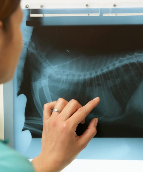
Digital X-Ray: Modern Diagnostic Imaging
X-ray, or radiology, is a vital diagnostic tool for imaging soft tissues, internal organs, and bones. Veterinary radiology has seen considerable advancements, and Caring Hands Animal Hospital utilizes state-of-the-art digital radiography.
Our digital radiology system offers the following benefits:
- Improved image resolution and manipulation, resulting in fewer attempts to obtain the best image.
- Lowered radiation exposure for your pet and our team.
- Reduced time required for radiograph procedures, lessening the need for sedation.
- High-resolution images that can be immediately emailed to specialists or saved on a CD for your records.
- Quicker diagnosis, allowing us to begin treatment sooner.
- Eco-friendly practices: eliminating film and processing chemicals.
Ultrasound Imaging for Pets
Ultrasound Imaging offered through our IMED department at our sister hospital Clarendon, or referral to local specialty hospital.
Ultrasound, or sonography, uses sound waves emitted from a probe that are transformed into visual images. These images are displayed on a screen, providing the veterinarian with a two-dimensional view of your pet's internal organs. Ultrasound allows the veterinarian to visualize and examine various organs and systems, including the heart, liver, gallbladder, spleen, kidneys, pancreas, and bladder.
Often, combining digital radiographs (X-rays) and ultrasound provides the most comprehensive evaluation of your pet's condition. Digital X-rays reveal the size, shape, and position of organs, while ultrasound allows the veterinarian to see the internal structure of these organs. Ultrasound is a minimally invasive and generally well-tolerated procedure for most pets.
Sometimes, the hair on the abdomen is clipped to improve image clarity during the ultrasound. A gel is then applied to the abdomen, and the ultrasound probe is moved systematically over the surface to capture images of each organ.
Because ultrasound provides real-time imaging, findings are immediately available. In some cases, the ultrasound images may be sent to a veterinary radiologist for further evaluation. When this occurs, the final report may take a few days.
Anesthesia is typically not required for ultrasound examinations. The procedure is completely painless, and most dogs and cats remain comfortable on a soft blanket during the scan. However, a sedative may be necessary for extremely anxious or uncooperative pets.
Its utility in pregnancy diagnosis, internal organ evaluation, and heart function assessment makes ultrasound an invaluable, non-invasive diagnostic tool for safeguarding your pet's health.