Ultrasound

An ultrasound, or sonogram, uses sound waves from a probe that are converted into an image. These images are displayed on the monitor
, giving the veterinarian a 2-dimensional “picture” of your pet’s organs. Ultrasound allows the veterinarian to visualize and examine many body organs and systems, including the heart, liver, gall bladder, spleen, kidney, pancreas and bladder.
Many times both digital radiographs (X-rays) and ultrasound of your pet are recommended for best evaluation of your pet’s problem. Digital X-rays show the size, shape and position of the organs and the ultrasound allows your veterinarian to see the internal structure of the organs. Ultrasound is very non-invasive and well tolerated by most pets.
The hair on the abdomen is sometimes clipped to allow adequate visualization during ultrasound. Gel is then placed on the abdomen and the ultrasound probe is moved methodically over the surface of the abdomen to record images of each organ.
Since an ultrasound study is performed in real time, the results of what is seen are known immediately. In some cases, the ultrasound images may be sent to a veterinary radiologist for further consultation. When this occurs, the final report may not be available for a few days.
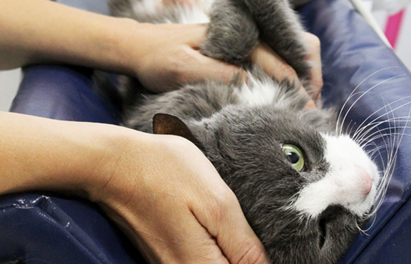
Anesthesia is not usually needed for most ultrasound examinations. The technique is totally painless and most dogs and cats will lie comfortably on a soft blanket while the scan is being performed. Occasionally, if the dog is very frightened or fractious, a sedative may be necessary.
Its usefulness for pregnancy diagnosis, evaluation of the internal organs, and assessment of heart function, make it an invaluable, non-invasive diagnostic tool to help protect to your pet’s well-being.
Radiology

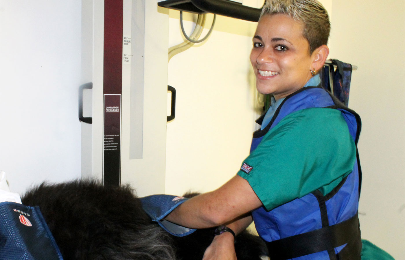
Radiology (or “x-ray”) is an invaluable diagnostic tool used to image soft-tissue structures, the internal organs, and bones.
Radiology has
come a long way in veterinary medicine over the past few years, and Caring Hands Animal Hospital is happy to offer state-of-the-art digital radiology.
The benefits of our digital radiology system include:
- Increased resolution and image manipulation abilities, which means fewer tries to get the best image.
- Decreased risk of exposure to your pet and our staff.
- Decreased time required to complete radiographs (and less sedation for pets/images that require it).
- High-quality images that may be instantly emailed to local specialists when indicated, and placed on a CD for your convenience
- Faster turn-around time allowing us to more quickly diagnose problems and begin the treatment process.
- Environmental safety: no more film or dangerous chemicals for processing.


